Bone Modeling From CT Scans
"3D-DOCTOR Software has been one of the tremendous analysis software that I use on a regular bases to extract information from image files to create 3D model.", A. Udoh, R&D Design Systems
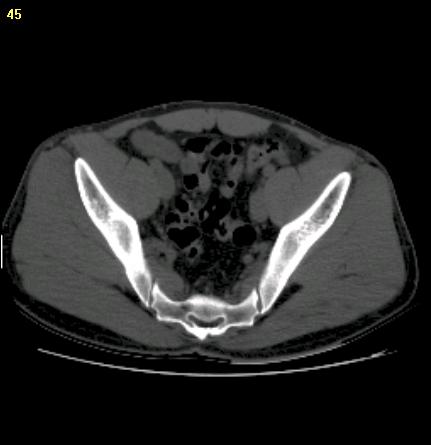
Step 1. Open Image |
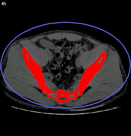
Step 2. Interactive Segmentation |
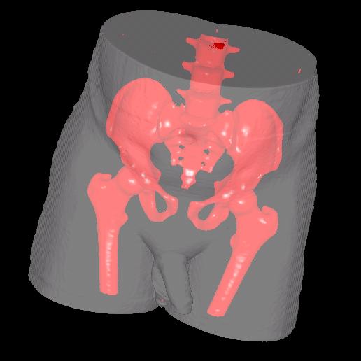
Step 3. Creating 3D Model |
Computed tomography (CT) is an imaging technique that uses special x-ray equipment to obtain cross-sectional images of the body. A CT image normally has different pixel intensity range for tissues such as bones, organs and other tissues. The threshold-based "Interactive Segmentation" provides an easy way to segment a CT image for 3D modeling.
A 3D mesh model can be created from a CT image in 3 main steps:
Step 1. Open the CT image. If the image slices come in as separate files, use the "New Stack" command.
Step 2. Use the "Interactive Segmentation" to generate object boundaries. For small size soft tissues, the manual tracing method can also be used. Boundaries can be edited using the boundary editor.
Step 3. Create 3D mesh models using the surface rendering command. The models can be exported to STL (ASCII and Binary), DXF, VRML, 3DS, OBJ, PLY and other formats for 3D measurement, rapid prototyping, simulation, treatment planning and other applications.